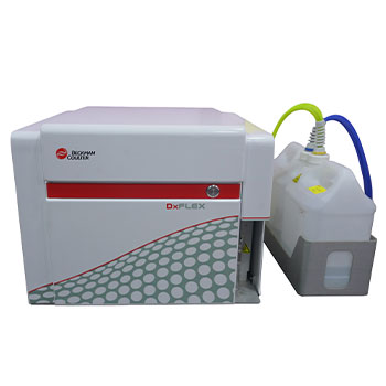
Flow Cytometry
Immunophenotyping is a test used to identify cells on the basis of the types of markers or antigens present on the cell’s surface, nucleus, or cytoplasm. This technique helps identify the lineage of cells using antibodies that detect markers or antigens on the cells. While some antigens are found only on one type of cell, others are found on different types. This process is widely used to diagnose different types of lymphoma and leukemia by comparing normal cells and cancer cells. It has become a common technique for the identification and classification of acute leukemia’s, particularly acute myeloid leukemia (AML).
Uses of Immunophenotyping
Immunophenotyping is widely used for the following reasons:
1) To differentiate between:
- Acute myeloid and lymphoid leukemia
- B and T cell lymphoid neoplasms such as chronic lymphocytic leukemia and lymphoma
- Reactive and neoplastic expansions of lymphocytes
2) Predicting prognosis in lymphoma
3) Identification of lymphocyte subsets

Flow cytometry is used for immunophenotyping.The sample may range from blood, fluids in the body cavity (such as peritoneal or pleural fluids), bone marrow, or solid tissues in liquid media.
Flow Cytometry
Flow cytometry is a technique used to analyze cells like cell counting, phenotyping, cell cycle assessment, and viability. Flow cytometry is a powerful tool which allows simultaneous multiparametric analysis of the physical and chemical characteristics of up to thousands of particles per second. This makes it a rapid and quantitative method for analysis and purification of cells in suspension. Using flow, one can determine the phenotype and function of the cells.
Applications:
- Analysis of whole human blood for diagnosing cancer initiating cells
- Immuno-phenotypic analysis
- Characterization and isolation of rare cells types like adult stem cells and
- Sorting different blood cell fractions for transplantations
Flow cytometry is generally used to determine cell lineage in leukemia and lymphoma.
Flow lymphoma is used in the case of lymphoid neoplasms or when a lymphoid origin is suspected on the basis of cell morphology after staining. This technique helps in prognostication and is also used to differentiate between neoplastic and reactive expansions of lymphocytes
Flow leukemia can be used in the case of an extensive range of leukemias that could be myeloid or lymphoid.
Flow Cytometry services overview:
1. Cell surface markers for AML, ALL (T-ALL, Pre B-ALL):
CD56, CD13, CD34, CD19, CD10, CD38, CD20, CD64, CD33, CD11c, CD117, CD11b, CD14, HLADR
Cytoplasmic markers for AML, ALL (T-ALL, Pre B-ALL):
MPO, CD79a, CD3, TdT
2. Secondary markers for AML-M6 and AML-M7
CD235a, CD36
3. Secondary markers for T-ALL
CD4, CD5, CD10, CD8, CD3,CD1a, CD2
4. Markers for Lymphoma (T-cell, B-cell)
CD23, CD10, CD79a, CD19, CD200, CD20, LAMBDA, KAPPA, CD5, CD19, CD3, CD38, CD7, CD4, CD8
5. Secondary markers for Burkitt ’s lymphoma
LAMBDA, KAPPA, CD20
6. Secondary markers for B-NHL
CD25, CD11c, CD19, CD123, CD45, CD19
7. Secondary markers for T-NHL
CD16, CD56, CD25
8. Markers for Paroxysmal Nocturnal Hemaglobinuria (PNH)
FLAER, CD59, CD14, CD45, CD24, CD64
9. Minimal Residual Disease (MRD) Markers for AML
CD45, CD34, CD117, HLADR, CD64, CD14, CD33, CD11B, CD7, CD19, CD56, CD38, CD123, CD36, CD15
10. MRD Markers for B cell ALL
CD20, CD10, CD19, CD45, CD34, CD58, CD38, CD123, CD200, CD73
11. MRD Markers for T cell ALL
CD3, CD7, CD45, CD34, CD56, CD38, CD5, CD4, CD8
12. MRD markers for Multiple Myeloma
CD38, CD138, CD19, CD45, CD56, CD117, KAPPA, LAMBDA
